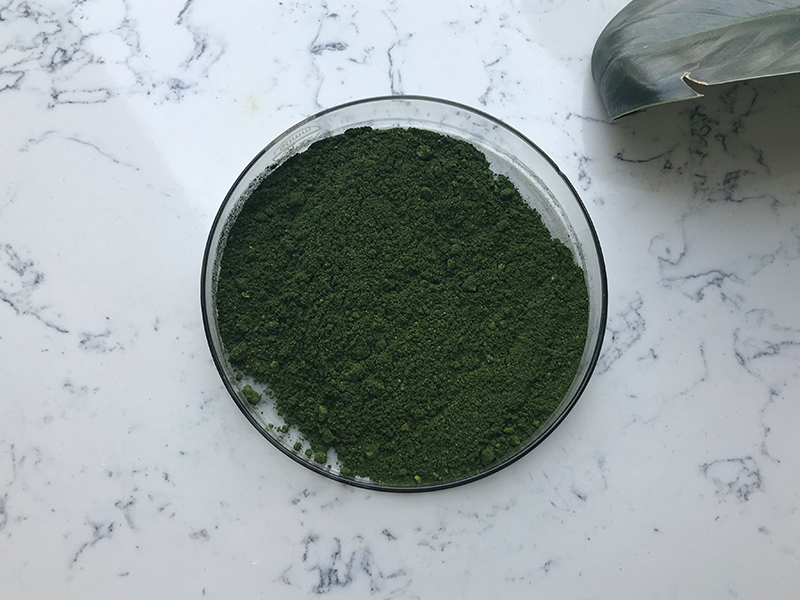
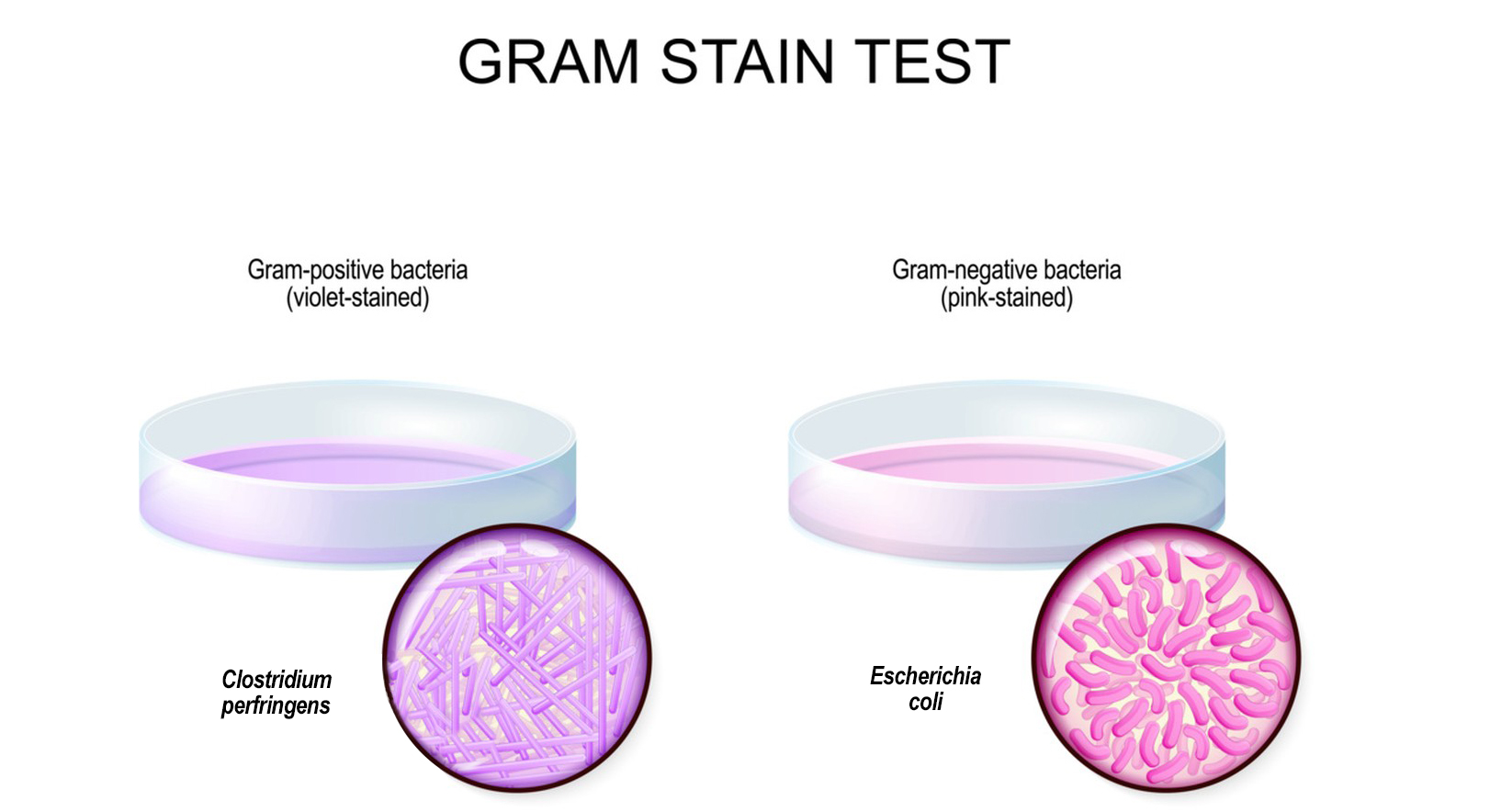
Gentian Violet are both dyes that are used for various purposes, including staining in microbiology and histology. Here are general guidelines for using crystal violet or gentian violet for staining:
Materials Needed:
1.Crystal violet or gentian violet solution
2.Microscope slides or other suitable surfaces for staining
3.Microscope coverslips (if applicable)
4.Gram’s iodine solution (for Gram staining, if applicable)
5.Ethanol or acetone (for destaining, if applicable)
6.Microscope

Procedure:
1.Preparation of Smear:
Prepare a thin smear of the sample you want to stain on a clean microscope slide.
Air-dry the smear to fix the cells to the slide.
2.Application of Gentian Violet:
Flood the smear with crystal violet or gentian violet solution. Ensure that the entire smear is covered.
Allow the dye to sit on the smear for a specific amount of time (this may vary depending on the staining protocol you are following).
3.Rinsing:
Rinse off the excess dye with water. This step is crucial to remove unbound dye.
4.Optional Gram’s Iodine Step (for Gram Staining):
If you are performing Gram staining, apply Gram’s iodine solution to the smear. This helps in fixing the dye to the cells.
5.Destaining (if applicable):
In some staining procedures, a destaining step is required to remove excess stain from the background. This is often done using ethanol or acetone.
6.Final Rinse:
Rinse the smear again with water to remove any remaining excess dye or destaining agent.
7.Drying:
Allow the slide to air-dry.
8.Mounting (if applicable):
If you are using coverslips, apply a mounting medium and gently place the coverslip over the stained smear.
9.Microscopic Examination:
Place the slide on a microscope stage and examine the stained cells under the microscope.

Notes:
Follow a staining protocol specific to the type of staining you are performing (e.g., Gram staining, simple staining).
Always handle dyes with care and follow safety guidelines.
The duration of staining, rinsing, and destaining steps may vary based on the staining protocol.
It’s important to note that specific protocols may vary depending on the type of staining you are performing and the nature of your sample. Always refer to a reliable staining protocol or laboratory manual for the most accurate and specific instructions.
