Phycocyanin in Cosmetics
Introduction to Phycocyanin
Phycocyanin, a primary active ingredient found in blue-green algae, has been the subject of numerous research studies due to its multifunctional properties. It is identified for its potent ability to scavenge oxygen radicals, while its rich array of amino acids lends it anti-aging and anti-allergenic qualities. This investigation delves into the feasibility of incorporating phycocyanin into the cosmetic industry, employing New Zealand white rabbits as an animal model to verify its skin toxicity through phototoxicity, acute percutaneous toxicity, and skin hypersensitivity tests.
Marine Algae: A Significant Natural Resource
Marine algae, an essential natural resource of the ocean, has gained considerable attention from researchers in recent years. Analysis of its chemical composition reveals that it contains a wealth of bioactive and nutritional substances, including polysaccharides, proteins, polyphenols, and sterols. Phycocyanin, also known as algal blue, is primarily found in blue-green algae and is a natural nutrient. With a molecular weight of approximately 30kD, it is an efficient light-capturing pigment protein and provides energy for the photosynthesis of algae.

The Multifunctional Attributes of Phycocyanin
Research has established that phycocyanin has a multitude of biological functions, including anti-coagulant, anti-inflammatory, immune-regulating, antioxidant, anti-allergic, and anti-tumoral properties. Moreover, due to its high water solubility, non-toxic nature, good coloring ability, and abundant readily absorbable amino acids, phycocyanin has been widely used in food colorants, health products, and animal feeds.
Utilization of Algae in Cosmetics
Given the non-toxic nature of algae and its rich nutrient content, it has increasingly been applied in the field of cosmetics in recent years. Similarly, algal red protein, a natural pigment, has been incorporated into the formulation of cosmetics for the development of natural pink and purple lipsticks and eyeliners. However, research into the use of phycocyanin in cosmetics has been relatively scarce. Studies have found that natural phycocyanin can eliminate hydroxyl and H2O2 radicals, demonstrating anti-aging effects. Since the promotion of aging by free radicals is a fact acknowledged by many researchers, natural antioxidants like phycocyanin present a broad application prospect as anti-aging active ingredients in cosmetics.
Investigating the Feasibility of Phycocyanin in Cosmetics
This study, based on the New Zealand white rabbit model, applies phycocyanin as a cosmetic on the skin surface and measures its skin toxicity through phototoxicity, acute percutaneous toxicity, and skin hypersensitivity experiments. The aim is to explore the feasibility of the natural antioxidant – phycocyanin for cosmetic development, providing a theoretical basis for the further development of new type-specific or compound non-toxic anti-aging beauty products.

Detailed Overview of Materials and Methods Used in the Study
1.1 Animals and Reagents
The subjects of this study are 66 healthy, qualified New Zealand white rabbits, half male and half female, with weights ranging from 2.6 to 2.8 kg. The animals are purchased from Yantai Raphael Biotechnology Co., Ltd. They are fed regular feed, have free access to water, and are kept in conditions with a temperature of 20-26°C, a daily temperature difference of no more than 4°C, and relative humidity of 40-70%. Illumination is controlled by a timer switch, with 12 hours of light and 12 hours of darkness, and the tray is replaced daily. Each rabbit is housed in a stainless steel cage. To help the animals adapt to their environment, they are housed under these conditions for five days before the experiment. During the experiment, a dosage of 2000mg/kg is administered. After the experiment, the animals are euthanized.
Three reagents are used in this study: phycocyanin, pure water, and cinnamaldehyde. Phycocyanin (purity of 3.2, represented as A620/A280) is purchased from Shin Daizo Spirulina Co., Ltd. Pure water is sourced from Hangzhou Wahaha Group Co., Ltd. Cinnamaldehyde is purchased from Guangdong Yongjiang Chemical Reagent Co., Ltd.
Comprehensive Experimental Approach for Equipment and Testing Methodologies
1.2 Instrumentation and Equipment
The instruments and equipment required for this study includes a Philips depilator and an Aiko UV irradiator for animal model preparation. The tissue embedding process uses a Leica EG1150 tissue embedding machine from Germany, while the slicing is done using a Leica rm2235 slicer, also from Germany. A VIP6 Sakura tissue dehydrator from Japan is used for tissue dehydration, and an Olympus microscope CCD imaging system (DP21), also from Japan, is used to observe and capture images of pathological slices.

1.3 Experimental Procedures and Testing
Various tests are conducted as part of the experiment, each one elucidating different aspects of the product’s effects.
1.3.1 Phototoxicity Test
In the phototoxicity test, six New Zealand white rabbits, half male and half female, are prepared. The skin on both sides of the animals’ spines is depilated 18-24 hours before the phototoxicity test, ensuring that the skin in the test area is intact, with no injuries or abnormalities. Four depilated areas are prepared, each with a depilated surface area of about 2cm x 2cm. After the animals are fixed, 0.2mL of the test substance is applied to depilated areas 1 and 2. This is a single-dose treatment, applied percutaneously. 30 minutes later, the left side (depilated areas 1 and 3) is covered with aluminium foil and secured with tape, while the right side undergoes UV irradiation. Skin reactions are observed at 1, 24, 48, and 72 hours post-irradiation, and each animal’s skin reaction score is determined based on Table 1 (Lin Jingyin, 2010).
1.3.2 Acute Percutaneous Toxicity Test
The acute percutaneous toxicity test involves thirty New Zealand white rabbits. They are randomly divided into three groups (ten individuals per group): a blank control group, a solvent control group, and a phycocyanin group. During the test, the daily observation indicators, water consumption, food consumption, weight and growth rate, and mortality rate of all groups are recorded. The hair on the back of the animals is removed 24 hours before the start of the test, exposing an area of skin approximately 10cm x 16cm for the intended poisoning area. The blank control group only undergoes depilation, the solvent control group is sprayed with pure water, and the phycocyanin group is uniformly coated with the test substance at a dose of 2000mg/kg. The phycocyanin is mixed with pure water to form a spreadable gel, which is uniformly applied to the back skin of the poisoning area, covered with thin film, and secured with non-irritating adhesive tape. After 24 hours, the poisoning ends, the residual test substance is removed with pure water, and observation and recording are carried out for 14 days. After the observation period, dissection is carried out, and pathological histological analysis is performed on the skin of the application site.
1.3.3 Skin Hypersensitivity Test
In the skin hypersensitivity test, thirty New Zealand white rabbits are again used. They are randomly divided into three groups (ten individuals per group): a blank control group, a cinnamaldehyde control group, and a phycocyanin group. The left side of the back hair is removed, exposing an area of skin about 10cm x 8cm for the intended poisoning area. The cinnamaldehyde control group is sprayed with 1% cinnamaldehyde, while the phycocyanin group is uniformly applied with a dose of 2000mg/kg.
1.3.4 Data Processing
For data analysis, the SPSS software is used to compare differences between groups. A t-test is used to judge whether the differences are significant. The results are represented as mean ± standard deviation, and a P value of less than 0.05 is considered statistically significant.
Results and Analysis
2.1 Phototoxicity
The phototoxicity of the test substance is evaluated by observing skin reactions in the subjects. When there is no skin reaction in the non-illuminated areas, and skin reactions with a total score of ≥2 are observed in at least one animal in the illuminated areas following application of the test substance, the substance is deemed to have phototoxicity. Our statistical results reveal that no significant phototoxic responses were observed in New Zealand white rabbits at a dosage of 2000mg/kg of phycocyanin. This suggests that when phycocyanin is used as a cosmetic topical, it does not exhibit phototoxicity.

2.2 Acute Dermal Toxicity
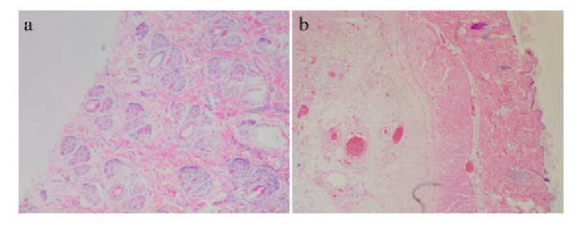
Throughout the duration of the experiment, none of the animals in the various groups exhibited signs of death, and no notable abnormalities were observed in terms of drinking water and food intake. Comparatively, no statistical difference was seen in the weight and weight growth rate of rabbits in the phycocyanin group compared to the control group, as shown in Table 2. Post-experiment dissection of the rabbits revealed no observable abnormalities in any organs. Further histopathological examinations were performed on tissue samples from the areas of administration in each group, revealing no significant abnormalities (Figure 2).
2.3 Skin Sensitization
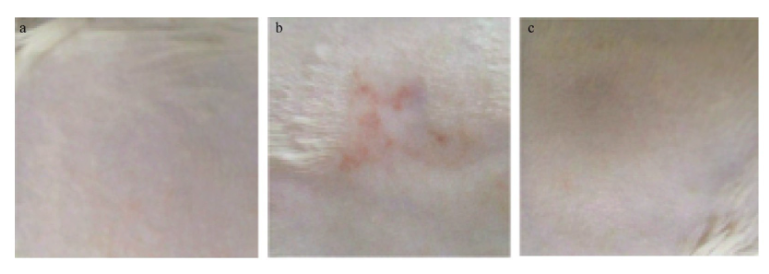
During the skin sensitization test, the weight and weight growth rate of animals in each group showed no significant statistical difference compared to the control group (Table 3). At a dosage of 2000mg/kg of phycocyanin, no skin sensitization reactions were observed in New Zealand white rabbits (Figure 3). However, the cinnamaldehyde control group significantly induced skin sensitization reactions in the rabbits. There were four animals with a skin sensitization score ≥2, accounting for 40% of the experimental group, indicating a moderate sensitization intensity. Both the phycocyanin group and the blank control group showed no sensitization reactions, with skin sensitization scores <2, and the difference compared to the cinnamaldehyde control group was significant (P<0.05) (Table 4).
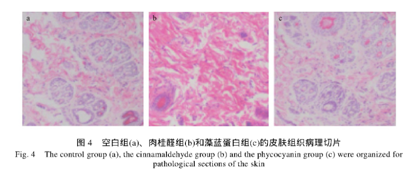
These results suggest that repeated exposure to phycocyanin does not cause allergic reactions such as erythema and edema in the skin of New Zealand white rabbits. Histopathological examinations were performed on tissue samples from the areas of administration in the blank group, the cinnamaldehyde control group, and the phycocyanin group. The results show that compared to the cinnamaldehyde control group, no notable abnormalities were observed in the phycocyanin group (Figure 4).
Discussion
3.1 Preliminary Feasibility of Phycocyanin as a Cosmetic
This experiment initiated a preliminary exploration into the feasibility of using phycocyanin as a cosmetic, testing its toxic effects on animal models. The research found that when phycocyanin was applied at a dosage of 2000mg/kg to the skin of New Zealand white rabbits, no notable phototoxicity, acute dermal toxicity, or skin sensitization effects were observed. However, whether these results can be extrapolated to the efficacy of human cosmetics still requires comprehensive evaluation in conjunction with other toxicity test results.
3.2 Antioxidant and Anti-aging Properties
Multiple studies have confirmed the antioxidant activity of phycocyanin and its ability to scavenge free radicals, attributing to it strong anti-aging effects. The human body’s defense system against oxidation includes enzymes such as superoxide dismutase (SOD) and glutathione peroxidase (GSHPX), which can fight oxidative stress responses and eliminate highly active free radicals produced in the body, thereby serving an anti-aging function. However, with aging, immune organs gradually atrophy, and the body’s immunity and anti-aging functions weaken. With its robust antioxidant capacity, phycocyanin may emerge as a new generation of anti-aging products.
3.3 Supporting Research
In 2010, Chu et al. extracted an aqueous solution containing phycocyanin from spirulina and studied its antioxidant capacity to scavenge free radicals using mouse cells. The results demonstrated that phycocyanin has a protective effect against free radical-induced cell apoptosis. Similarly, Niraj’s research group chose a nematode model to test the anti-aging effect of phycocyanin and its inhibitory effect on endogenous protein homeostasis (Singh et al., 2015). Research by Harishkumar et al. also explored the influence of phycocyanin on growth factors and cell migration in the wound healing process (Li et al., 2016).
Recently, Castangia et al. (2016) addressed the issue of bioavailability by successfully encapsulating phycocyanin in capsules. In vitro permeation experiments indicated that this method promotes deeper deposition of phycocyanin in the skin, thereby enhancing phycocyanin’s antioxidant activity against human keratinocytes. All these studies suggest the potential of phycocyanin as an antioxidant to resist aging both internally and externally.
Conclusion and Future Prospects
4.1 Can Phycocyanin Be Used in Cosmetics on a Large Scale?
In conclusion, whether phycocyanin can be massively applied in the beauty and cosmetics industry depends on whether it can meet strict quality control standards. In China, the development and utilization of algal proteins is still in its infancy, with most still at the laboratory research stage and application development just beginning. Therefore, the selection, isolation, extraction, and purification of algal species, the stability of active ingredients, the optimization of administration routes such as topical, oral, and injection, and determining whether the effective components of phycocyanin are its entirety or its metabolites are key areas of focus for future research.
Based on the aforementioned studies, phycocyanin has a broad application prospect in the cosmetics field. This promising future, coupled with the exciting results of ongoing research, marks an exciting time for the advancement of phycocyanin as a potential key ingredient in the world of cosmetics.
