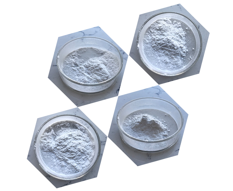
DMSA (Dimercaptosuccinic Acid) is a chelating agent primarily used for the treatment of heavy metal poisoning, particularly lead poisoning. It works by binding to heavy metals in the bloodstream and facilitating their excretion through the urine.
The main conditions DMSA is used to treat include:
- Lead Poisoning: DMSA is commonly used in children and adults to treat lead toxicity. It is considered a first-line treatment for moderate lead poisoning.
- Mercury Poisoning: DMSA can also be used to treat mercury poisoning, although it is not as commonly used for this purpose as for lead poisoning.
- Arsenic Poisoning: It may be used for arsenic poisoning in some cases, though other chelating agents (like dimercaprol) are often preferred for severe cases.
- Other Heavy Metal Toxicities: DMSA has been studied for its use in treating poisoning by other metals such as cadmium and thallium, but these indications are less common.
DMSA is typically administered orally and is considered to have a lower toxicity profile compared to other chelating agents, making it a preferred option for outpatient treatment.

Inspection method of DMSA
DMSA (Dimercaptosuccinic Acid) is a radiopharmaceutical used in a renal scan, typically for evaluating renal function and structure. The inspection or imaging method using DMSA is commonly called a DMSA renal scan or renal scintigraphy. Here’s an outline of how the method works:
1. Preparation:
- Patient Preparation: There is usually no special preparation needed before a DMSA scan, though patients may be asked to drink plenty of fluids or avoid medications that affect kidney function.
- Radioactive Injection: A small dose of DMSA, labeled with a radioactive isotope (usually Technetium-99m), is injected intravenously. The DMSA binds to the renal tubules in the kidneys.
2. Mechanism:
- Once injected, the radiolabeled DMSA is filtered by the kidneys and is taken up by the renal cortical tissue. The radioactivity from the DMSA is detected by a gamma camera that captures images of the kidney’s structure and function.
3. Imaging Process:
- After injection, the patient typically needs to wait 2-3 hours to allow for optimal uptake of the radiopharmaceutical by the kidneys.
- The patient is then positioned under a gamma camera, which takes images of the kidneys from different angles.
- The images obtained allow for the evaluation of renal cortical function, structure, and perfusion.
4. Evaluation:
- The images show the distribution of the radiopharmaceutical in the kidneys. Abnormalities such as scarring, asymmetry in kidney size or function, or reduced uptake can indicate conditions like pyelonephritis, glomerulonephritis, renal scarring, or chronic kidney disease.
- The renal cortical scarring is commonly assessed, as it provides information about past infections or injuries to the kidney tissue.
5. Advantages:
- Provides functional and structural information about the kidneys.
- Non-invasive and can help detect early kidney damage.
- Useful in conditions such as chronic pyelonephritis, vesicoureteral reflux, and renal scars.

6. Limitations:
- The technique may not be as effective in assessing renal function in the presence of acute renal failure or other more diffuse kidney disorders.
- Exposure to a small amount of radiation, although the dose is generally low and considered safe for diagnostic imaging purposes.
In summary, the DMSA scan is a sensitive test for assessing renal function and detecting abnormalities in the kidney structure, particularly for conditions that cause cortical scarring or other chronic damage.
